8Protein Structure Determination
A protein is a chain of amino acids that folds into one specific three-dimensional shape, and that shape is most of what determines what the protein does. For fifty years, learning the shape meant running a hard, slow experiment. The load-bearing idea of this chapter is that the folded coordinates are, to a startling degree, predictable from the sequence alone, because evolution leaves a statistical fingerprint of which residues sit next to each other in space. Modern predictors read that fingerprint and return atomic coordinates in seconds instead of months. But a predicted structure is a single snapshot, and a snapshot is not the whole biology. The temptation, after 2021, is to say structure is "solved." It is not. It is predicted well for many proteins, which is a different and more useful claim.
8.1The problem: from sequence to 3D
The sequence of a protein is one-dimensional: a string over an alphabet of twenty amino acids, which you met in the genetics primer (Chapter 5). The function lives in three dimensions. Two residues far apart in the string can be neighbors in the folded shape, forming the pocket that binds a drug or the cleft that cuts a bond. Going from the string to the coordinates is the protein folding problem, and for most of its history the only reliable route was experiment.
Those experiments are the ground truth, and they are expensive. X-ray crystallography (measuring how a crystal of the protein diffracts X-rays) can resolve atoms sharply but first demands that the protein crystallize, which many proteins simply refuse to do. Cryo-electron microscopy, or cryo-EM (imaging flash-frozen molecules with an electron beam), skips crystals and now handles large machines and membrane proteins, and it has grown to roughly a third of new depositions. Both feed the Protein Data Bank (PDB), the open archive of experimentally determined structures introduced in the data chapter (Chapter 4) (Berman et al., 2000). The PDB holds on the order of 200,000 structures, which sounds like plenty until you compare it with the hundreds of millions of known sequences. That gap is the whole opportunity.
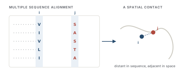
What makes prediction possible is co-evolution. Line up many evolutionary relatives of a protein into a multiple sequence alignment (MSA) (a stack of homologous sequences, one per row, with corresponding positions in columns; a homolog is a related sequence from another species or gene copy). If two positions are in physical contact, a mutation at one that would disrupt the pair tends to be compensated by a mutation at the other. Across enough relatives, those two columns change together. Contact leaves a correlation, and correlation is something a model can learn to read.
Intuition
A structure predictor is a decoder for a code evolution already wrote: which columns of an alignment vary in lockstep tells you which residues are neighbors in space.
Collaborator
"If it just needs an alignment, what happens to a protein with no known relatives?" The co-evolution signal weakens, and MSA-based accuracy drops. Orphan proteins, fast-evolving viral proteins, and designed sequences are exactly where you should distrust a confident-looking prediction, and where single-sequence models (below) become interesting rather than merely faster.
8.2Models that attempt it
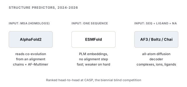
The breakthrough was AlphaFold2 (Jumper et al., 2021). Its input is an MSA plus any known template structures; its network reasons jointly over the alignment and a map of pairwise residue relationships, then folds that into coordinates. At CASP14 in 2020, the biennial blind competition where predictors get sequences whose structures are withheld, AlphaFold2 reached accuracy competitive with experiment for a large fraction of targets. That result is what reset the field. It also ships a calibrated confidence score, pLDDT, per residue, and low pLDDT is your first warning that a region is unreliable or disordered.
AlphaFold2 predicts a single protein chain (a monomer). Real biology is assemblies: proteins bound to other proteins, to DNA and RNA, to small-molecule ligands and metal ions. AlphaFold3 (Abramson et al., 2024) rebuilds the output end as a diffusion model (a generator that starts from noise and iteratively denoises toward a structure, the same family you will meet again in protein design, Chapter 9). Working at the level of individual atoms rather than only amino-acid residues lets one unified network predict complexes: protein–ligand, protein–nucleic-acid, ion-bound. It beats specialized docking tools at their own tasks. RoseTTAFold All-Atom (Krishna et al., 2024) reaches the same generality by a different route, mixing a residue-level representation for the protein backbone with an atom-level one for everything else.
A second axis is how much evolutionary context the model demands. ESMFold (Lin et al., 2023) drops the MSA entirely: it reads structure off the internal representations of ESM-2, a protein language model (PLM) trained on tens of millions of raw sequences (roughly 65 million unique UniRef sequences) (the PLM idea from Chapter 4). One sequence in, coordinates out. It trades some accuracy on hard targets for a large speedup and no alignment step, which makes it the tool of choice for scanning millions of sequences or for proteins with no relatives to align.
The 2024–2026 shift is that AlphaFold3-class capability is no longer a single lab's. Open-weight co-folding models (predicting the joint structure of a complex, sequence and ligand folded together) now match it closely: Boltz-1 (MIT) and Boltz-2 (MIT and Recursion) (Wohlwend et al., 2024; Passaro et al., 2025) and Chai-1 from Chai Discovery (Chai Discovery, 2024) both reproduce AlphaFold3-level accuracy under open licenses, and Boltz-2 adds a binding-affinity head that outperformed every method submitted to the CASP16 affinity challenge in a retrospective evaluation. For an ML practitioner, this matters: you can run and fine-tune these yourself.
Collaborator
"Which one should my lab actually run?" If you have relatives and want the single best monomer, AlphaFold2 is still a strong default. If you need a complex with a ligand, nucleic acid, or ion, reach for an all-atom co-folder (AlphaFold3, Boltz, Chai, or RoseTTAFold All-Atom). If you are screening millions of orphan sequences or want it fully in-house, ESMFold or an open co-folder wins on speed and control.
Note
Do not read a benchmark headline as a promise about your protein. CASP scores are averages over a target set; a model near the top can still be wrong on a specific membrane protein, a large assembly, or a region with few homologs. Always check the per-residue confidence.
8.3What they do well and what is still hard
Be precise about the win. For a single, well-behaved protein with plenty of evolutionary relatives, these models return a fold that is often as good as an experimental one, in seconds, for free. That genuinely changes how biology gets done. The honest boundary of the win is just as important.
The deepest limitation is that the models return one static structure, and a protein is not one shape. Most proteins flex between multiple functional states, an open and a closed form, a bound and an unbound one, and the biology often lives in the motion, not any single frame. AlphaFold was trained to predict the single most probable structure, so it collapses a conformational ensemble (the population of shapes a molecule actually visits) down to its dominant mode (Jing et al., 2024). Active work now coaxes ensembles out of these networks, by injecting experimental data or by pairing the predictor with flow-matching generators, but returning a faithful distribution of states rather than a point estimate remains open.
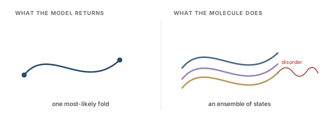
Related failure modes cluster here. Intrinsically disordered regions (IDRs), stretches that have no fixed fold at all and stay floppy in the cell, are common and functional, and a model that outputs coordinates will draw some shape anyway, flagged only by low confidence. Large multi-component assemblies stress the models more than single chains. And a prediction that depends on a strong MSA degrades exactly where relatives are scarce.
Common trap
A confident-looking ribbon diagram is not the same as understanding function. The model gives you coordinates; it does not tell you which pocket is the active site, how tightly a drug binds, or how the shape changes when it does its job. Structure is an input to those questions, not an answer to them. Binding affinity and function are separate prediction problems (property prediction, Chapter 7; design, Chapter 9).
Collaborator
"So can I skip the experiment now?" For a first pass, often yes, and that saves real time. But when the answer must be right, a mechanism claim, a drug program, a novel fold with no close relatives, you still validate against experiment. The models are a powerful prior over structure, not a replacement for measuring it.
The mental model to carry forward: sequence-to-structure went from a grand-challenge problem to a fast, reliable first draft for most proteins. The frontier moved to what a single frame cannot express: dynamics, ensembles, disorder, and the leap from a shape to the function that shape performs.
References
- Abramson, J., Adler, J., Dunger, J., Evans, R., Green, T., Pritzel, A., Ronneberger, O., Willmore, L., et al. (2024). Accurate structure prediction of biomolecular interactions with AlphaFold 3. Nature.
- Berman, H. M., Westbrook, J., Feng, Z., Gilliland, G., Bhat, T. N., Weissig, H., Shindyalov, I. N., & Bourne, P. E. (2000). The Protein Data Bank. Nucleic Acids Research.
- Chai Discovery (2024). Chai-1: Decoding the molecular interactions of life. bioRxiv.
- Jing, B., Berger, B., & Jaakkola, T. (2024). AlphaFold Meets Flow Matching for Generating Protein Ensembles. ICML. arXiv:2402.04845.
- Jumper, J., Evans, R., Pritzel, A., Green, T., Figurnov, M., Ronneberger, O., Tunyasuvunakool, K., Bates, R., et al. (2021). Highly accurate protein structure prediction with AlphaFold. Nature.
- Krishna, R., Wang, J., Ahern, W., Sturmfels, P., Venkatesh, P., Kalvet, I., Lee, G. R., Baker, D., et al. (2024). Generalized biomolecular modeling and design with RoseTTAFold All-Atom. Science.
- Lin, Z., Akin, H., Rao, R., Hie, B., Zhu, Z., Lu, W., Smetanin, N., Verkuil, R., et al. (2023). Evolutionary-scale prediction of atomic-level protein structure with a language model. Science.
- Passaro, S., Corso, G., Wohlwend, J., Reveiz, M., Thaler, S., Somnath, V. R., Getz, N., Portnoi, T., et al. (2025). Boltz-2: Towards Accurate and Efficient Binding Affinity Prediction. bioRxiv.
- Wohlwend, J., Corso, G., Passaro, S., Getz, N., Reveiz, M., Leidal, K., Swiderski, W., Atkinson, L., et al. (2024). Boltz-1: Democratizing Biomolecular Interaction Modeling. bioRxiv.
Check yourself
Questions a sharp collaborator might ask on this chapter. Pick an answer to see whether it holds up.
-
A structure predictor gives a confident, sharp fold for a well-studied enzyme but a low-confidence, tangled result for a newly discovered orphan protein with no known relatives. What is the most likely reason?
MSA-based predictors depend on co-evolution across many relatives. With few homologs, the alignment carries little contact signal and accuracy drops. This is exactly the regime where single-sequence models like ESMFold become attractive, though they too lose accuracy on genuinely hard, relative-poor targets. -
Why does AlphaFold2 alone struggle to model a protein bound to a small-molecule drug and a magnesium ion, and what changed with AlphaFold3?
AlphaFold2 is a monomer/protein predictor. AlphaFold3's key architectural change is an atom-level diffusion generator that represents and folds arbitrary chemical components together, letting one network handle complexes and beat specialized docking tools. Open weights are a separate story that belongs to Boltz and Chai. -
A collaborator says 'AlphaFold solved protein structure, so we can stop doing crystallography.' What is the sharpest correction?
The models return one static structure and collapse the population of states a molecule visits. Dynamics, intrinsic disorder, large assemblies, and the leap from shape to function all remain open, which is why 'solved' overstates the case even though the first-draft fold is often excellent. -
What does a per-residue pLDDT confidence score most usefully flag when it is very low over a contiguous stretch of a prediction?
A model that outputs coordinates will place atoms even for a floppy, fold-free region; low pLDDT is your signal that the drawn shape is untrustworthy there. Disordered regions are common and functional, so a confident-looking ribbon over a low-pLDDT stretch is a classic way to be misled. -
By 2025-2026, what is the most accurate statement about open-weight structure models like Boltz and Chai relative to AlphaFold3?
The 2024-2026 shift is that co-folding capability is no longer confined to one lab: open models reproduce AlphaFold3-level accuracy on complexes under permissive licenses, and Boltz-2 even added a binding-affinity head that outperformed every method submitted to the CASP16 affinity challenge in a retrospective evaluation. For a practitioner, in-house runnability and fine-tuning are the practical payoff.